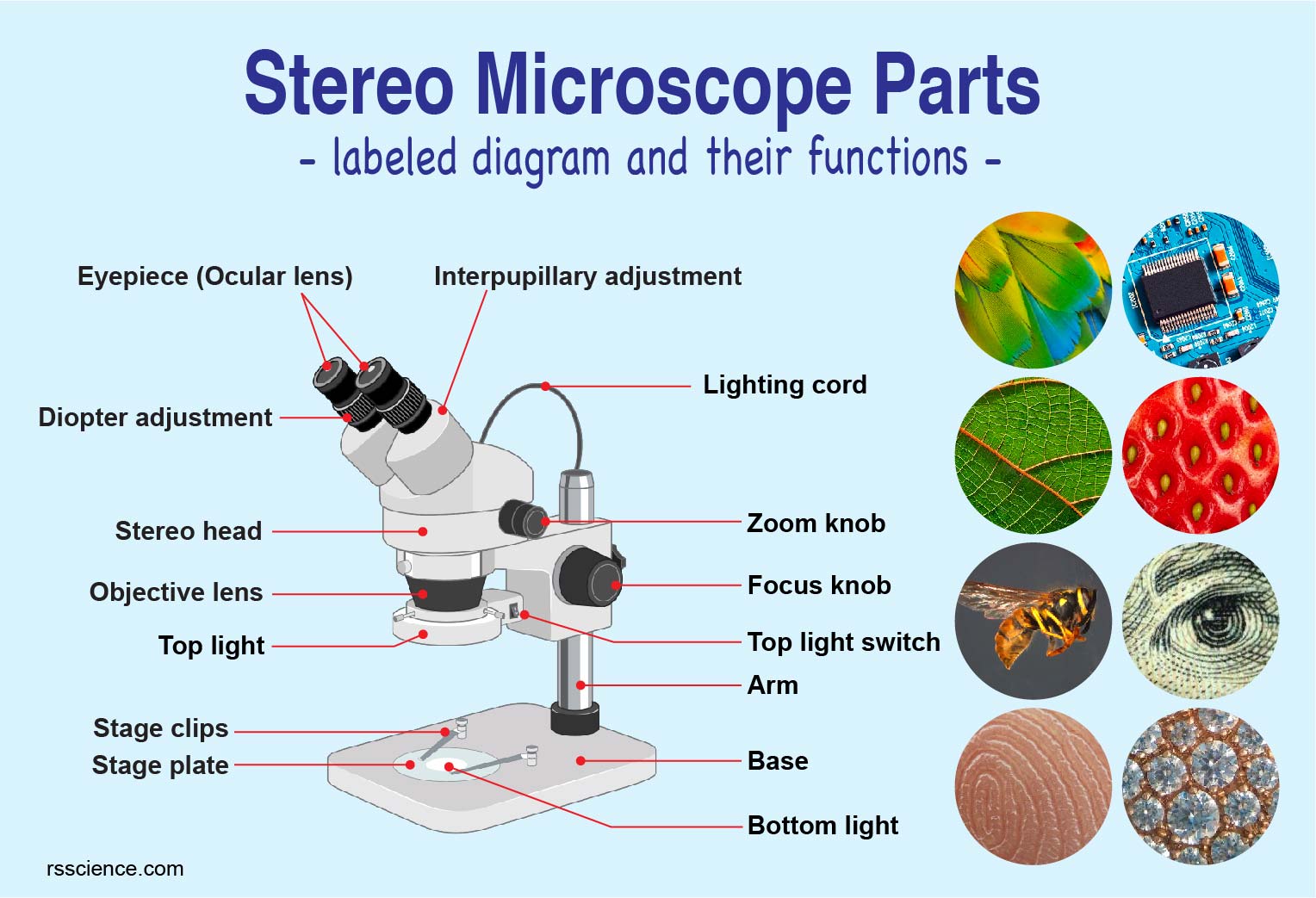
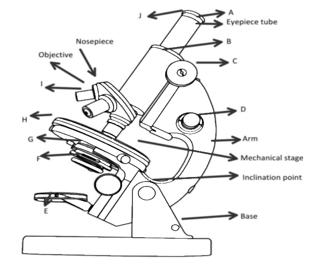
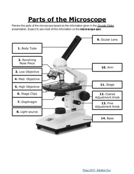
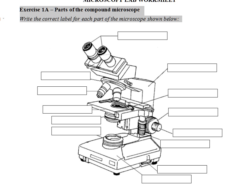
45 pictures of microscope with label
Swollen Dog Paw and Toe Bumps: Pictures and Tips - Dog Health Guide More is not better, and may seriously harm or kill your dog. Typically, for a swollen dog paw, a canine patient is prescribed a dose of 5 to 10 mg per pound, 2x per day. Note that children's aspirin is 81 mg. (check the label). Only use after checking with your veterinarian. Your vet is in the best position to judge the length of treatment and ... 467,408 Microscope Images, Stock Photos & Vectors | Shutterstock 467,408 microscope stock photos, vectors, and illustrations are available royalty-free. See microscope stock video clips Image type Orientation Color People Artists More Sort by Popular Science College and University Healthcare and Medical Jobs/Professions Biology microscope laboratory scientist medicine lens Next of 4,675
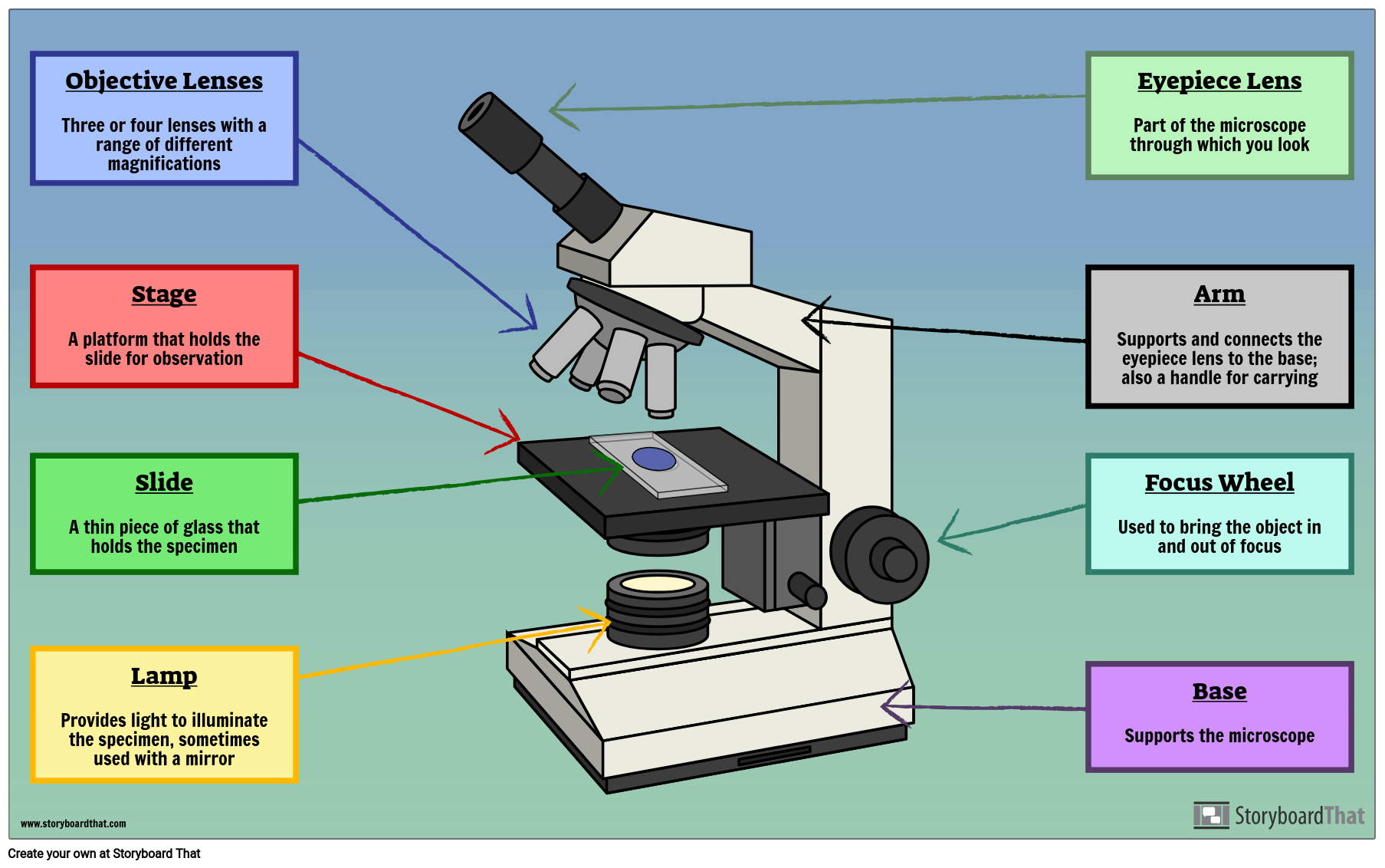
Parts of a Microscope Labeling Activity - Storyboard That In this activity, students will create a poster of a microscope with labeled parts. Students will identify and describe the microscope parts and functions. This is an awesome activity to complete at the beginning of either the school year or the unit on basic cells. ... All storyboards and images are private and secure. Teachers can view all of ...
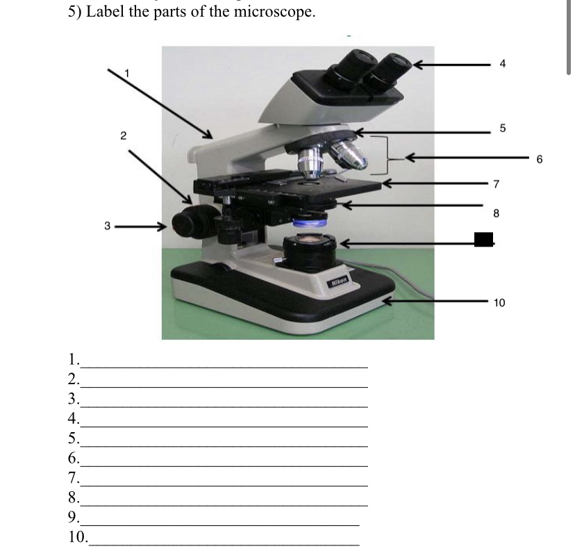
Pictures of microscope with label
scienceexplorers.com › guide-to-teaching-kidsGuide to Teaching Kids About Cells | Science Explorers Apr 25, 2019 · Give each student a chance to look through the microscope at the cells. Point out that each slide contains numerous cells. Repeat the process with the second slide. Have students draw pictures of what they saw under the microscope and guess what the cells do. Finish with an explanation of the cell and its organelle functions. Microscope Parts, Function, & Labeled Diagram - slidingmotion Microscope Parts Labeled Diagram The principle of the Microscope gives you an exact reason to use it. It works on the 3 principles. Magnification Resolving Power Numerical Aperture. Parts of Microscope Head Base Arm Eyepiece Lens Eyepiece Tube Objective Lenses Nose Piece Adjustment Knobs Stage Aperture Microscopic Illuminator Condenser Lens Parts of the Microscope with Labeling (also Free Printouts) Parts of the Microscope with Labeling (also Free Printouts) By Editorial Team March 7, 2022 A microscope is one of the invaluable tools in the laboratory setting. It is used to observe things that cannot be seen by the naked eye. Table of Contents 1. Eyepiece 2. Body tube/Head 3. Turret/Nose piece 4. Objective lenses 5. Knobs (fine and coarse) 6.
Pictures of microscope with label. There’s No Scientific Basis for Race—It's a Made-Up Label There’s No Scientific Basis for Race—It's a Made-Up Label It's been used to define and separate people for millennia. But the concept of race is not grounded in genetics. By Elizabeth Kolbert. photographs by Robin Hammond. Published 22 Oct 2018, 16:32 BST. Skulls from the collection of Samuel Morton, the father of scientific racism, illustrate his classification of people into five … PDF Parts of a Microscope Printables - Homeschool Creations Label the parts of the microscope. You can use the word bank below to fill in the blanks or cut and paste the words at the bottom. Microscope Created by Jolanthe @ HomeschoolCreations.net. Parts of a eyepiece arm stageclips nosepiece focusing knobs illuminator stage objective lenses › people-and-cultureThere’s No Scientific Basis for Race—It's a Made-Up Label “They’re cute, aren’t they?” she says, opening the cage to show me. The mice look ordinary, with sleek brown coats and shiny black eyes. But examined under a microscope, they are different from their equally cute cousins in subtle yet significant ways. 1.6 Skill: Identifying stages of mitosis under a microscope and on a ... This video takes you through microscope images of cells going through mitosis and identifies the different phases under the microscope and on a micrograph.
Wipers - Wikipedia Wipers was a punk rock band formed in Portland, Oregon, in 1977 by guitarist and vocalist Greg Sage, along with drummer Sam Henry and bassist Dave Koupal. The group's tight song structure and use of heavy distortion were hailed as extremely influential by numerous critics and musicians. They are also considered to be the first Pacific Northwest punk band. 18,889 Microscope slide Images, Stock Photos & Vectors - Shutterstock Microscope slide royalty-free images. 18,889 microscope slide stock photos, vectors, and illustrations are available royalty-free. See microscope slide stock video clips. Image type. Animal Cell Anatomy & Diagram - Enchanted Learning The cell is the basic unit of life. All organisms are made up of cells (or in some cases, a single cell). Most cells are very small; in fact, most are invisible without using a microscope. Cells are covered by a cell membrane and come in many different shapes. The contents of a cell are called the protoplasm. Glossary of Animal Cell Terms: Cell ... www1.udel.edu › biology › ketchamUD Virtual Compound Microscope - University of Delaware ©University of Delaware. This work is licensed under a Creative Commons Attribution-NonCommercial-NoDerivs 2.5 License.Creative Commons Attribution-NonCommercial-NoDerivs 2
› top-30-most-common-lab-equipmentTop 30 Most Common Lab Equipment Names, Pictures And Their ... Jan 17, 2022 · Light microscope: It uses lights and a series of magnifying lenses to observe a tiny specimen; Dark‐field microscope: It is used to observe live spirochetes, such as those that cause syphilis. Phase‐contrast microscope: It is an optical microscopy technique that converts phase shifts in the light passing through a transparent specimen to ... Rocks: Pictures of Igneous, Metamorphic and Sedimentary … A Grain of Sand Gallery of sand grains through a microscope by Dr. Gary Greenberg. Puddingstone. Puddingstone - a conglomerate with clasts that contrast in color with the matrix. Alamy Images. Geology Dictionary. Geology Dictionary - contains thousands of geological terms with their definitions. Difficult Rocks . Difficult Rocks Elementary students find lots of rocks that … Top 30 Most Common Lab Equipment Names, Pictures And Their … 17/01/2022 · Below are some of the common lab equipment pictures, their names, and their various uses in the lab. Measuring beakers ; Beakers are one of the common laboratory apparatus. They are flat bottom glassware or plasticware used to store, heat, and mix substances. They vary in size and are also used to hold liquids during titration and filtration processes. … #1 Victor Traps | eBay Nous voudrions effectuer une description ici mais le site que vous consultez ne nous en laisse pas la possibilité.
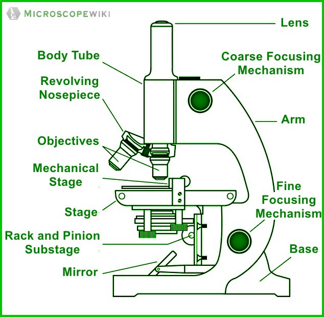
Microscope Parts and Functions Microscope Parts and Functions With Labeled Diagram and Functions How does a Compound Microscope Work?. Before exploring microscope parts and functions, you should probably understand that the compound light microscope is more complicated than just a microscope with more than one lens.. First, the purpose of a microscope is to magnify a small object or to magnify the fine details of a larger ...
300+ Free Microscope & Laboratory Images - Pixabay 399 Free images of Microscope Related Images: laboratory science bacteria research scientist lab biology chemistry medical Find your perfect microscope image. Free pictures to download and use in your next project.
Label the microscope — Science Learning Hub Label the microscope Interactive Add to collection Use this interactive to identify and label the main parts of a microscope. Drag and drop the text labels onto the microscope diagram. coarse focus adjustment diaphragm or iris stage eye piece lens fine focus adjustment base high-power objective light source Download Exercise Tweet
UD Virtual Compound Microscope - University of Delaware ©University of Delaware. This work is licensed under a Creative Commons Attribution-NonCommercial-NoDerivs 2.5 License.Creative Commons Attribution-NonCommercial-NoDerivs 2.5 License.
en.wikipedia.org › wiki › Microscope_image_processingMicroscope image processing - Wikipedia Microscope image processing is a broad term that covers the use of digital image processing techniques to process, analyze and present images obtained from a microscope. Such processing is now commonplace in a number of diverse fields such as medicine, biological research, cancer research, drug testing, metallurgy, etc. A number of ...
geology.com › rocksRocks: Pictures of Igneous, Metamorphic and Sedimentary Rocks Photographs and information for a large collection of igneous, metamorphic and sedimentary rocks. Geology.com
Microscope Labeled Pictures, Images and Stock Photos View microscope labeled videos Browse 49 microscope labeled stock photos and images available, or start a new search to explore more stock photos and images. Newest results Fluorescent Imaging immunofluorescence of cancer cells growing... Microscope diagram vector illustration. Labeled zoom instrument... Microscope diagram vector illustration.
Microscope Stock Photos, Pictures & Royalty-Free Images - iStock Browse 196,265 microscope stock photos and images available, or search for magnifying glass or microscope isolated to find more great stock photos and pictures. Newest results magnifying glass microscope isolated science microscope icon laboratory scientist microscope coronavirus microscope electron microscope bacteria microscope lab microscope
› swollen-dog-pawSwollen Dog Paw and Toe Bumps: Pictures and Tips More is not better, and may seriously harm or kill your dog. Typically, for a swollen dog paw, a canine patient is prescribed a dose of 5 to 10 mg per pound, 2x per day. Note that children's aspirin is 81 mg. (check the label). Only use after checking with your veterinarian.
Guide to Teaching Kids About Cells | Science Explorers 25/04/2019 · Have students draw pictures of what they saw under the microscope and guess what the cells do. Finish with an explanation of the cell and its organelle functions. Ask the kids how big the largest cell is. Answer by showing them an ostrich egg, which is the largest single cell and 10,000 times larger than the cheek cells they saw. Give kids unlabeled pictures of plant …
Microscope Labeling Game - PurposeGames.com About this Quiz. This is an online quiz called Microscope Labeling Game. There is a printable worksheet available for download here so you can take the quiz with pen and paper. This quiz has tags. Click on the tags below to find other quizzes on the same subject. Science.
18,701 Microscope drawing Images, Stock Photos & Vectors - Shutterstock Find Microscope drawing stock images in HD and millions of other royalty-free stock photos, illustrations and vectors in the Shutterstock collection. Thousands of new, high-quality pictures added every day.
Connective Tissue Images Labeled | Virtual Anatomy Lab VAL - ncccval Epithelium Images Labeled. Epithelium Images Unlabeled. Connective Tissue Images Labeled. Connective Tissue Images Unlabeled. Microscope. Microscope Images Labeled. Microscope Images Unlabeled. Mitosis. Mitosis Images Labeled.
Binocular Microscope Pictures, Images and Stock Photos Browse 814 binocular microscope stock photos and images available, or search for microtome or histology to find more great stock photos and pictures. Newest results microtome histology cryostat optical devices in vector format equipment and science experiments Trinocular microscope on blue background Cartoon illustrated image of a microscope
Microscope image processing - Wikipedia Microscope image processing is a broad term that covers the use of digital image processing techniques to process, analyze and present images obtained from a microscope.Such processing is now commonplace in a number of diverse fields such as medicine, biological research, cancer research, drug testing, metallurgy, etc.A number of manufacturers of …
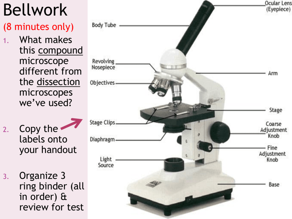
Parts of a microscope with functions and labeled diagram - Microbe Notes Q. List down the 18 parts of a Microscope. 1. Ocular Lens (Eye Piece) 2. Diopter Adjustment 3. Head 4. Nose Piece 5. Objective Lens 6. Arm (Carrying Handle) 7. Mechanical Stage 8. Stage Clip 9. Aperture 10. Diaphragm 11. Condenser 12. Coarse Adjustment 13. Fine Adjustment 14. Illuminator (Light Source) 15. Stage Controls 16. Base 17.
PDF Label parts of the Microscope Label parts of the Microscope: . Created Date: 20150715115425Z
Compound Microscope: Definition, Diagram, Parts, Uses, Working ... - BYJUS A compound microscope is defined as. A microscope with a high resolution and uses two sets of lenses providing a 2-dimensional image of the sample. The term compound refers to the usage of more than one lens in the microscope. Also, the compound microscope is one of the types of optical microscopes. The other type of optical microscope is a ...
Compound Microscope Parts - Labeled Diagram and their Functions The eyepiece (or ocular lens) is the lens part at the top of a microscope that the viewer looks through. The standard eyepiece has a magnification of 10x. You may exchange with an optional eyepiece ranging from 5x - 30x. [In this figure] The structure inside an eyepiece. The current design of the eyepiece is no longer a single convex lens.
Parts of the Microscope with Labeling (also Free Printouts) Parts of the Microscope with Labeling (also Free Printouts) By Editorial Team March 7, 2022 A microscope is one of the invaluable tools in the laboratory setting. It is used to observe things that cannot be seen by the naked eye. Table of Contents 1. Eyepiece 2. Body tube/Head 3. Turret/Nose piece 4. Objective lenses 5. Knobs (fine and coarse) 6.
Microscope Parts, Function, & Labeled Diagram - slidingmotion Microscope Parts Labeled Diagram The principle of the Microscope gives you an exact reason to use it. It works on the 3 principles. Magnification Resolving Power Numerical Aperture. Parts of Microscope Head Base Arm Eyepiece Lens Eyepiece Tube Objective Lenses Nose Piece Adjustment Knobs Stage Aperture Microscopic Illuminator Condenser Lens
scienceexplorers.com › guide-to-teaching-kidsGuide to Teaching Kids About Cells | Science Explorers Apr 25, 2019 · Give each student a chance to look through the microscope at the cells. Point out that each slide contains numerous cells. Repeat the process with the second slide. Have students draw pictures of what they saw under the microscope and guess what the cells do. Finish with an explanation of the cell and its organelle functions.











Post a Comment for "45 pictures of microscope with label"