45 abdominal muscles labeled
Abdominal Muscles Function, Anatomy & Diagram | Body Maps - Healthline The rectus abdominis is the large muscle in the mid-section of the abdomen. It enables the tilt of the pelvis and the curvature of the lower spine. Next to it on both sides of the body is the... human anatomy muscle abdominals Muscle anatomy muscles female exercise chart body abdominal lower human guide abdomen fitness groups workouts left posters drawing charts exercises. Anatomy muscle woman man alamy kneeling similar. ... abdominal cat muscles anatomy updated2 sunyorange bio edu. Cat Muscles Labeled | Specimens : Cat : Thorax , Abdomen , Medial Leg pinterest.com.
Abdominal Pain During Pregnancy: Causes and Treatment Braxton Hicks Contractions: Sometimes labeled “practice contractions,” Braxton Hicks is more of a mild annoyance than a risk to you or your baby. Many women report that Braxton Hicks feel like a tightening of the stomach muscles so your stomach feels firm or hard. It is important to differentiate Braxton Hicks from true contractions.
Abdominal muscles labeled
Atlas of CT Anatomy of the Abdomen - W-Radiology A computer develops separate images, also called slices, of the abdomen. Medical imagery produced from a CT scan may be stored, viewed on a computer monitor, or printed on film. Three-dimensional models of the abdomen may be created by stacking the slices together. An abdominal CT scan should take 15 to 30 minutes to perform (3). Abdominal Deep Muscles Anatomy & Diagram | Body Maps - Healthline The rectus abdominis is the large muscle in the middle portion of the abdomen. It facilitates the tilt of the pelvis and the curvature of the lower spine. Next to it on both sides of the body is... Abdominal wall: Layers, muscles and fascia | Kenhub Lateral flat muscle group situated on either side of the abdomen, which includes three muscles: external oblique, internal oblique and transversus abdominis . Anterior vertical muscles situated bilaterally to the median fibrous structure called linea alba. They are called rectus abdominis and pyramidalis muscles.
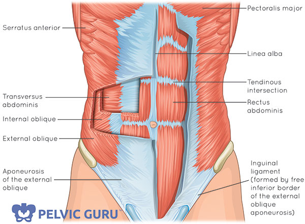
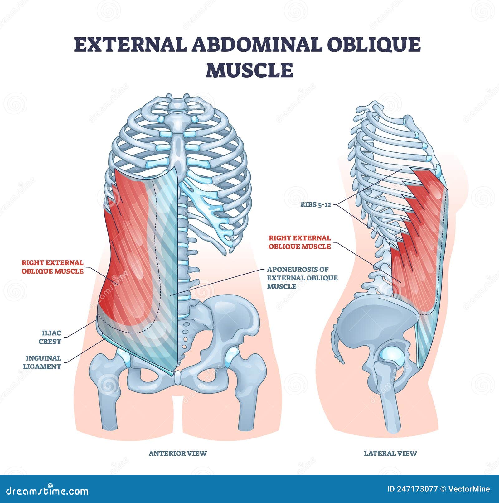
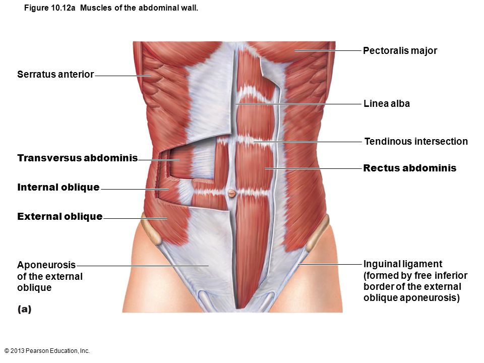
Abdominal muscles labeled. Meet Your Muscles: Abdominal Muscles Anatomy - Bodybuilding Wizard Abdominal Muscles Anatomy. The abdominal wall can be divided into two separate anatomic parts, each of which functions differently. The front wall consists of one muscle, the rectus abdominis (also known as the "abs"). The rectus abdominis starts near the middle of the sternum and runs vertically to below your navel. Muscles of the Abdomen - TeachMeAnatomy The anterolateral abdominal wall consists of four layers- skin, superficial fascia (connective tissue), muscles and parietal peritoneum. The muscles of the anterolateral abdominal wall include flat and vertical muscles. The flat muscles are stacked on top of each other and have fibres that run in different directions, helping to strengthen the ... Understanding the Human Stomach Anatomy With Labeled Diagrams Given below is a labeled diagram of the stomach to help you understand stomach anatomy. The stomach is divided into four parts. These include: Cardia Fundus Body Pylorus Cardia refers to the section of the stomach that is located around the cardiac orifice. The lower esophageal sphincter lies at the junction where the esophagus meets the stomach. Univ of Michigan - Gross Anatomy - Muscles of the Abdomen the inguinal ligament is a specialization of the external abdominal oblique aponeurosis; the external spermatic fascia is the external abdominal oblique muscle's contribution to the coverings of the testis and spermatic cord. thoracolumbar fascia, anterior 2/3 of the iliac crest, lateral 2/3 of the inguinal ligament.
UC San Diego's Practical Guide to Clinical Medicine The major components of the abdominal exam include: observation, auscultation, percussion, and palpation. While these are the same elements which make up the pulmonary and cardiac exams, they are performed here in a slightly different order (i.e. auscultation before percussion) and carry different degrees of importance. Learn all muscles with quizzes and labeled diagrams | Kenhub Apr 26, 2022 · The number of muscles of the body is estimated to be around 600, so there’s certainly a lot to cover. A good way to get started is to learn using a regional approach. For example, starting by learning the muscles of the upper extremity , then the muscles of the lower extremity, and so on. The Anatomy Of Your Abdominal Muscles - Caliber Fitness The rectus abdominis is positioned between the ribs and the pubic bone at the front of the pelvis, and is actually made up of 8 distinct muscle bellies. When the muscle contracts, these muscle bellies are visible, assuming low enough levels of body fat, creating that 'six-pack' look. Retroperitoneal space - Wikipedia Retroperitoneal structures. Structures that lie behind the peritoneum are termed "retroperitoneal". Organs that were once suspended within the abdominal cavity by mesentery but migrated posterior to the peritoneum during the course of embryogenesis to become retroperitoneal are considered to be secondarily retroperitoneal organs.
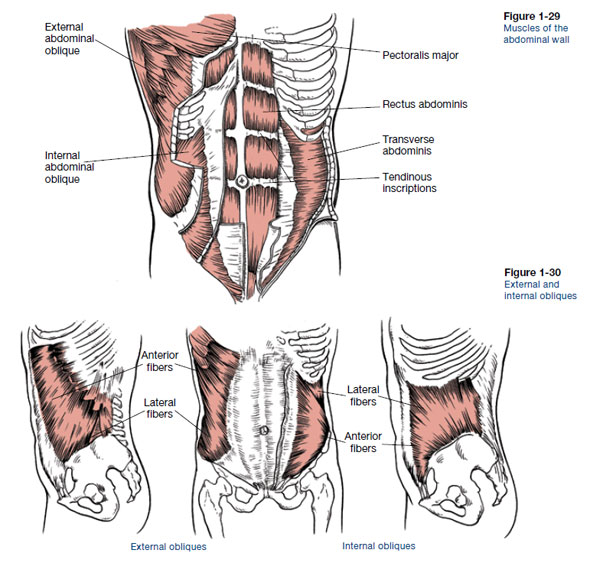
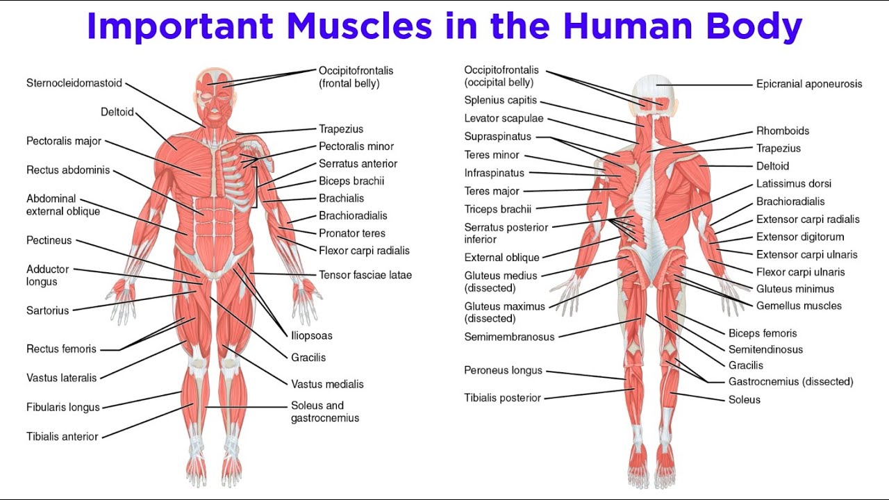
Abdominal muscles: Anatomy, Function, Exercise The abdominal muscles form the anterior & lateral abdominal wall & consist of the external abdominal obliques, the internal abdominal obliques, the rectus abdominis & the transversus abdominis. Acting together these muscles form a firm wall that protects the viscera & they help to maintain erect posture. Abdomen Anatomy, Area & Diagram | Body Maps - Healthline The major muscles of the abdomen include the rectus abdominis in front, the external obliques at the sides, and the latissimus dorsi muscles in the back. The major organs of the abdomen include the... Psoas major muscle - Wikipedia Psoas major labeled at bottom left. Diagram of a transverse section of the posterior abdominal wall, to show the disposition of the lumbodorsal fascia. Muscles of the iliac and anterior femoral regions. abdominal muscle | Description, Functions, & Facts | Britannica abdominal muscle, any of the muscles of the anterolateral walls of the abdominal cavity, composed of three flat muscular sheets, from without inward: external oblique, internal oblique, and transverse abdominis, supplemented in front on each side of the midline by rectus abdominis.
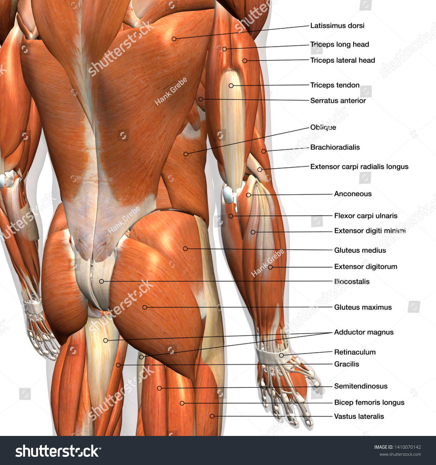
Ilustrasi Stok Labeled Anatomy Chart Male Back Muscles ...
Abdominal muscles: Anatomy, Function, Exercise The deep abdominal muscles, together with the intrinsic back muscles, make up the core muscles and help keep the body stable and balanced, and help to protect the spine. There are main four muscles in the abdomen: External Abdominal obliques I nternal Abdominal obliques Rectus abdominis Transversus abdominis External Abdominal obliques
Muscles – Advanced Anatomy 2nd. Ed.
Anatomy, Abdomen and Pelvis, Abdominal Wall - NCBI Bookshelf The abdominal muscles may be divided broadly into anterolateral and posterior components. Anterolateral muscles include five paired muscles: the external oblique, internal oblique, transversus abdominis, rectus abdominis, and pyramidalis. [2] Posterior muscles include psoas major and quadratus lumborum bilaterally.
Learn all muscles with quizzes and labeled diagrams | Kenhub
Muscles of the Abdominal Region | UAMS Department of Neurobiology and ... the inguinal ligament is a specialization of the external abdominal oblique aponeurosis; the external spermatic fascia is the external abdominal oblique muscle's contribution to the coverings of the testis and spermatic cord. oblique, internal abdominal. thoracolumbar fascia, anterior 2/3 of the iliac crest, lateral 2/3 of the inguinal ligament.
Abdominal Muscle Diagram Images – Browse 5,555 Stock Photos ...
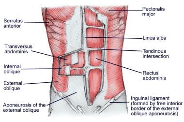
The Complete Guide to Your Abs Muscles - Shape First, a little anatomy lesson. Along with muscles in the lower back, these key abdominals make up your core. External Obliques: The outer layer of the abs on your sides; these run diagonally downward. Internal Obliques: Just underneath the external obliques, these run diagonally up your sides. Rectus Abdominis: Two paired sheets of muscle from ...

The Anatomy Of Your Abdominal Muscles
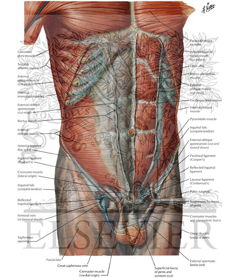
Abdomen (Anatomy): Definition, Function, Muscles - Biology Dictionary The abdominal muscles consist of three distinct layers residing within the abdominal wall and extend to the pubis, iliac crest, lower ribs, and vertebral column. The muscle fibers merge at the midline, surround the rectus abdominus, and join on the other side at a point known as the linea alba.

File:Muscles anterior labeled.png - Wikimedia Commons
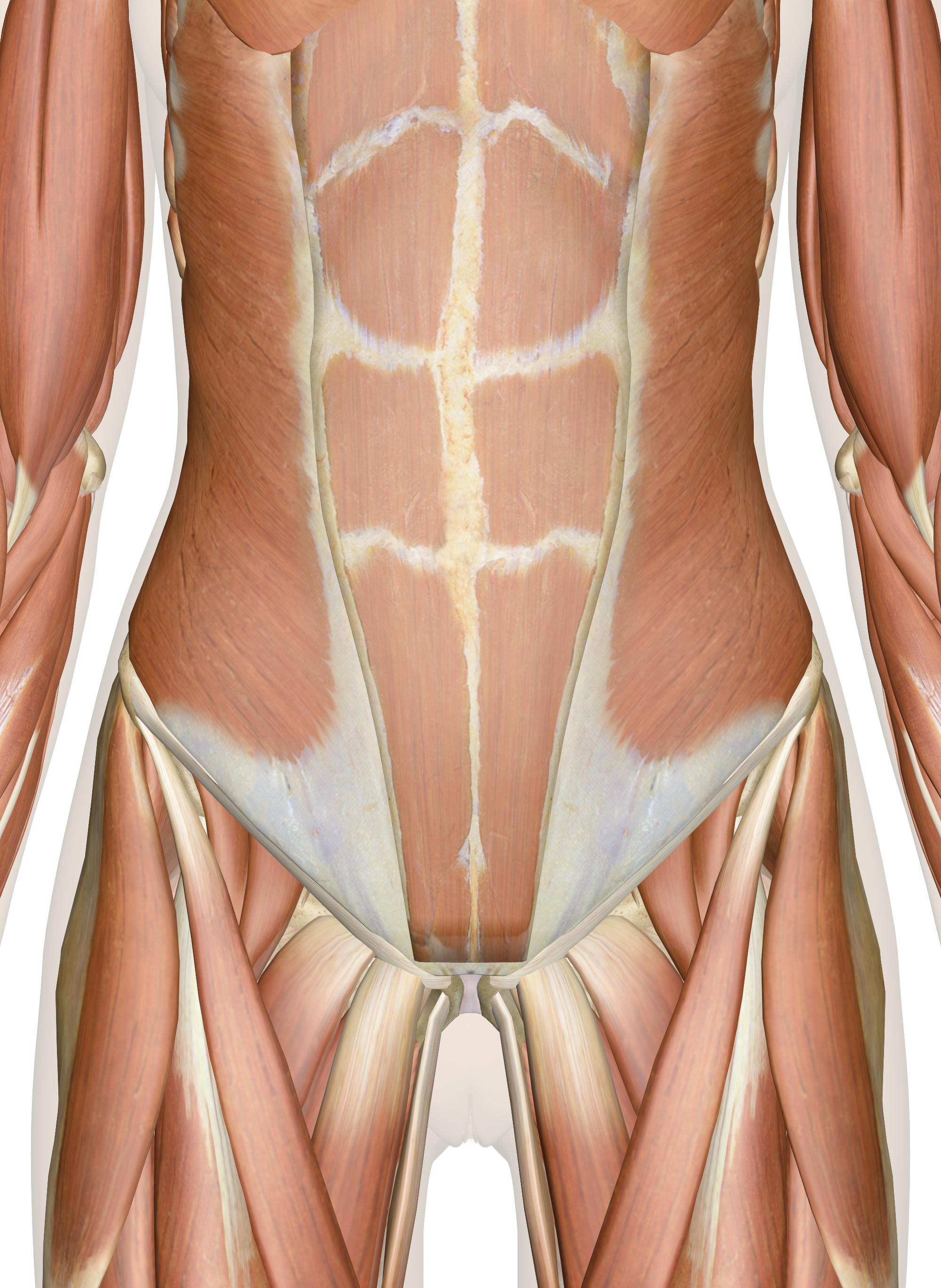
Meet Your Muscles: Abdominal Muscles Anatomy - Fitness Blog The two rectus abdominis muscles (one on each side) are encased in a sheath of fascia that forms the central demarcation down the middle of the abs, known as the linea alba. Fascia divisions in the muscles are responsible for the "six-pack" appearance. If you can see your linea alba right now, congratulations; you're leaner than we are.
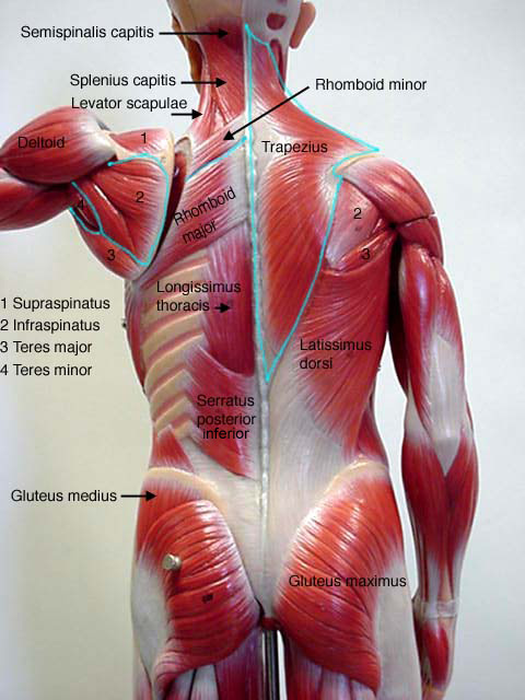
Male Muscle Model
Abdominal Muscles - Physiopedia The abdominal muscles are the muscles forming the abdominal walls, the abdomen being the portion of the trunk connecting the thorax and pelvis. An abdominal wall is formed of skin, fascia, and muscle and encases the abdominal cavity and viscera [1].

Anterior Muscles of the Human Body (labelled), illustration ...
400 Abdominal Muscles Anatomy Stock Photos - Dreamstime Browse 400 professional abdominal muscles anatomy stock photos available royalty-free. Sporty sexy girl with great abdominal muscles in a white t-shirt with beautiful Breasts drip drops of water on black backgroun. Seductive body. candid Breasts. Bodybuilder Abdominal Muscles. Bodybuilder with his hands clasped above his head displaying his ...

Abdominal muscles Diagram | Quizlet
My Stomach is Not Digesting Food Properly - Health Advisor Gastroparesis is a condition that occurs when the stomach takes too long to empty food. This condition, also referred to as postponed gastric emptying, is a result of unusual motion of the muscles in the stomach. 1. This can be due to either nerve damage or muscular disease.

Anatomy of the Female Abdomen and Pelvis, Cut-away View ...
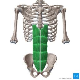
Abdominal Muscles Location and Function - Verywell Fit The most well-known and prominent abdominal muscle is the rectus abdominis. It is the long, flat muscle that extends vertically between the pubis and the fifth, sixth, and seventh ribs. The rectus abdominis connects to the xiphoid process, a bony landmark at the bottom of the sternum.
The Transverse Abdominals: Your Inner Corset - Your Pace Yoga
Abdominal Muscles • Muscles that act on the Abdomen • Anatomy & Function The abdominal wall is divided into the anterolateral and posterior wall. There are five sets of anterolateral abdominal muscles. From superficial to deep, they are: Pyramidalis muscle (present only in 80% of individuals) Rectus abdominis muscle External abdominal oblique muscle Internal abdominal oblique muscle Transverse abdominis muscle

Abdominal external oblique muscle - Wikiwand
Anterior abdominal muscles: Anatomy and functions | Kenhub The name 'rectus abdominis' means 'straight abdominal', and is indicative of the parallel direction the fibres of the muscle take as they pass from their origin to their point of insertion. The muscle has a strap-like shape, and is narrower inferiorly, widening as it passes superiorly. Rectus abdominis muscle Musculus rectus abdominis 1/3

WHAT ARE THE CORE MUSCLES? - The Core Expert
Abdominal Muscles: Anatomy and Function - Cleveland Clinic Your abdominal muscles have many important functions, from holding organs in place to supporting your body during movement. There are five main muscles: pyramidalis, rectus abdominus, external obliques, internal obliques, and transversus abdominis. Ab strains and hernias are common, but several strategies can keep your abs safe and healthy.

495 Rectus Abdominis Stock Photos, Pictures & Royalty-Free ...
Abdominal Muscles : Attachment, Nerve Supply & Action - Anatomy Info The four main abdominal muscle is described is given below: Transversus Abdominis The transversus abdominis muscle is the deepest of the abdominal muscles, lying internally to the internal abdominal obliques. Its main roles are to stabilize the trunk and maintain internal abdominal pressure. Origin
Muscles of the Abdomen, Lower Back and Pelvis
Anatomy of the Abdominal Wall and Core Muscles - Body Contouring There are four layers of abdominal muscles that we will talk about today: Rectus abdominis muscles, aka "six-pack" abs The most commonly known abdominal muscle group is the rectus abdominis. These are best known as the "six-pack" muscles. This is the most superficial abdominal muscle group because it lies closest to your skin.
Abdominal Muscles Function, Anatomy & Diagram | Body Maps
141,764 Abdominal muscles Images, Stock Photos & Vectors - Shutterstock 141,866 abdominal muscles stock photos, vectors, and illustrations are available royalty-free. See abdominal muscles stock video clips Image type Orientation Color People Artists More Sort by Popular Recreation/Fitness Biology Healthcare and Medical Anatomy muscle organ abdomen physical fitness bodybuilding human body gymnasium Next of 1,419

Muscles of trunk and abdomen - Stock Image - C020/0426 ...
Muscles of the Abdomen, Lower Back and Pelvis - Innerbody The abdominal muscles also play a major role in the posture and stability to the body and compress the organs of the abdominal cavity during various activities such as breathing and defecation. The muscles of the lower back, including the erector spinae and quadratus lumborum muscles, contract to extend and laterally bend the vertebral column.
Abdominal muscles anatomy Vector Art Stock Images | Depositphotos
Learn the facial muscles with quizzes & labeled diagrams - Kenhub Mar 03, 2022 · To begin, spend a few minutes analysing the face muscles diagram above in which all the face muscles are clearly labeled. Once you think you’ve got a solid idea of the location of each muscle (top tip: try to associate the location with the function to aid your memory!), you can try labelling the muscles yourself using our facial muscles ...

Muscles of the Neck and Torso - Classic Human Anatomy in ...
Abdominal wall: Layers, muscles and fascia | Kenhub Lateral flat muscle group situated on either side of the abdomen, which includes three muscles: external oblique, internal oblique and transversus abdominis . Anterior vertical muscles situated bilaterally to the median fibrous structure called linea alba. They are called rectus abdominis and pyramidalis muscles.

Lower Back Muscles Labeled Educational Anatomical Scheme ...
Abdominal Deep Muscles Anatomy & Diagram | Body Maps - Healthline The rectus abdominis is the large muscle in the middle portion of the abdomen. It facilitates the tilt of the pelvis and the curvature of the lower spine. Next to it on both sides of the body is...
Goldsgym.co.id - PT Fit and Health Indonesia
Atlas of CT Anatomy of the Abdomen - W-Radiology A computer develops separate images, also called slices, of the abdomen. Medical imagery produced from a CT scan may be stored, viewed on a computer monitor, or printed on film. Three-dimensional models of the abdomen may be created by stacking the slices together. An abdominal CT scan should take 15 to 30 minutes to perform (3).
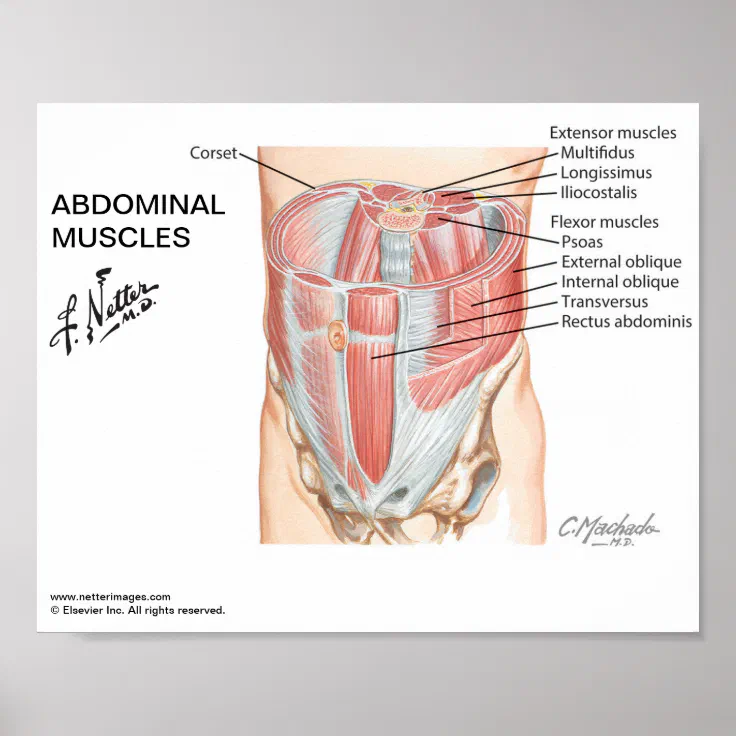
Small poster - Netter's Abdominal Muscles | Zazzle
Regions and Planes of the Abdomen: Overview, Abdominal Skin ...
:max_bytes(150000):strip_icc()/GettyImages-1192965863-2000-e1583791850721-ead103f609e84db993821e671abe1b1c.jpg)
The Complete Guide to Your Abs Muscles
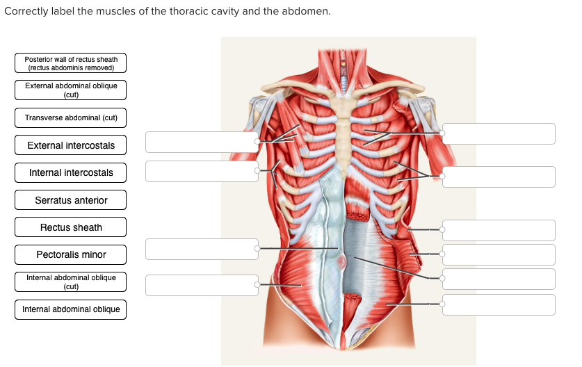
Solved Correctly label the muscles of the thoracic cavity ...
Multichoice The Muscular System Flashcards - Easy Notecards
Core Anatomy | Learn About Core Muscles | ACE Blog

Pectoral and abdominal muscles, illustration - Stock Image ...

Chest and abdominal muscles Diagram | Quizlet
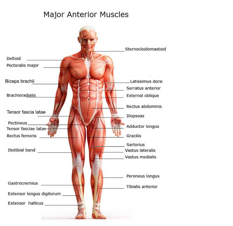
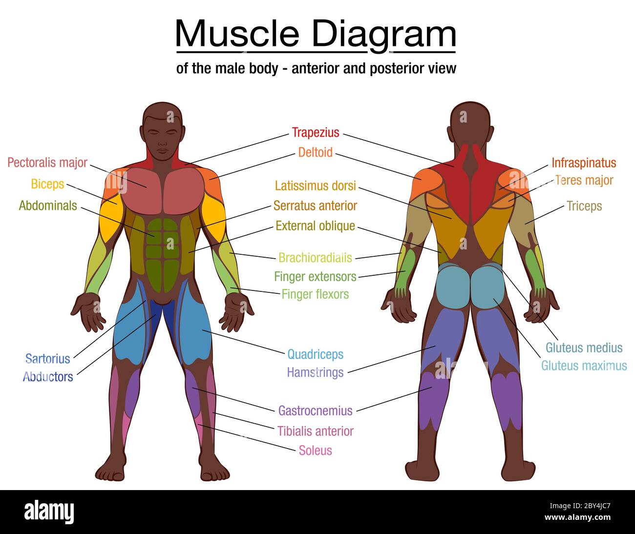
Chart of Major Muscles on the Front of the Body with Labels
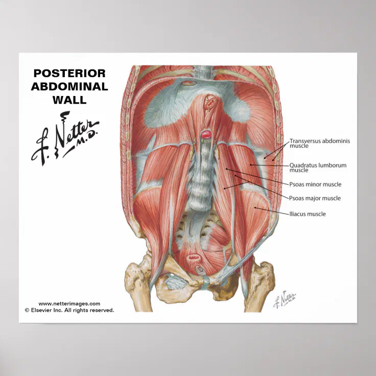
Netter's Posterior Abdominal Wall - Labeled Chart | Zazzle

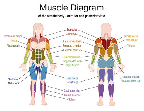
The Female Muscular System Laminated Anatomical Chart ...
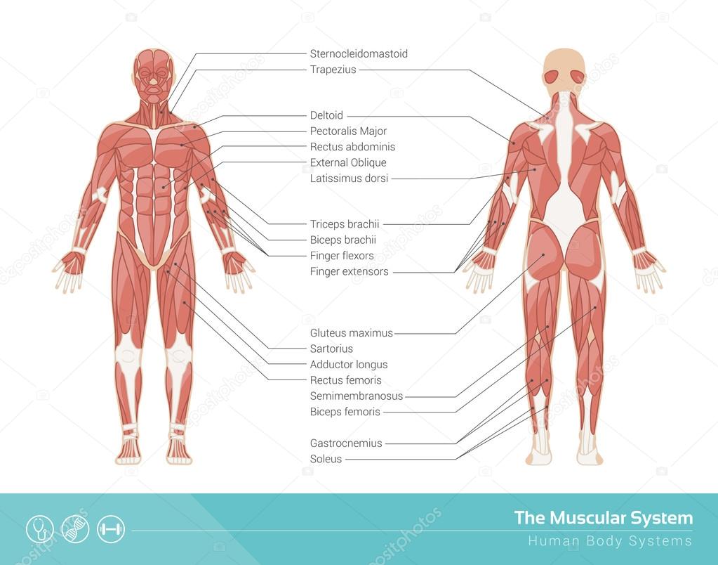
The Muscular System
Ilustrasi Stok Male Biceps Chest Muscle Chart Labeled ...
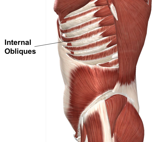
Oblique Muscle Stock Illustrations – 1,130 Oblique Muscle ...

Oblique Muscle - The Definitive Guide | Biology Dictionary
Anterior Abdominal Wall: Intermediate Dissection ...
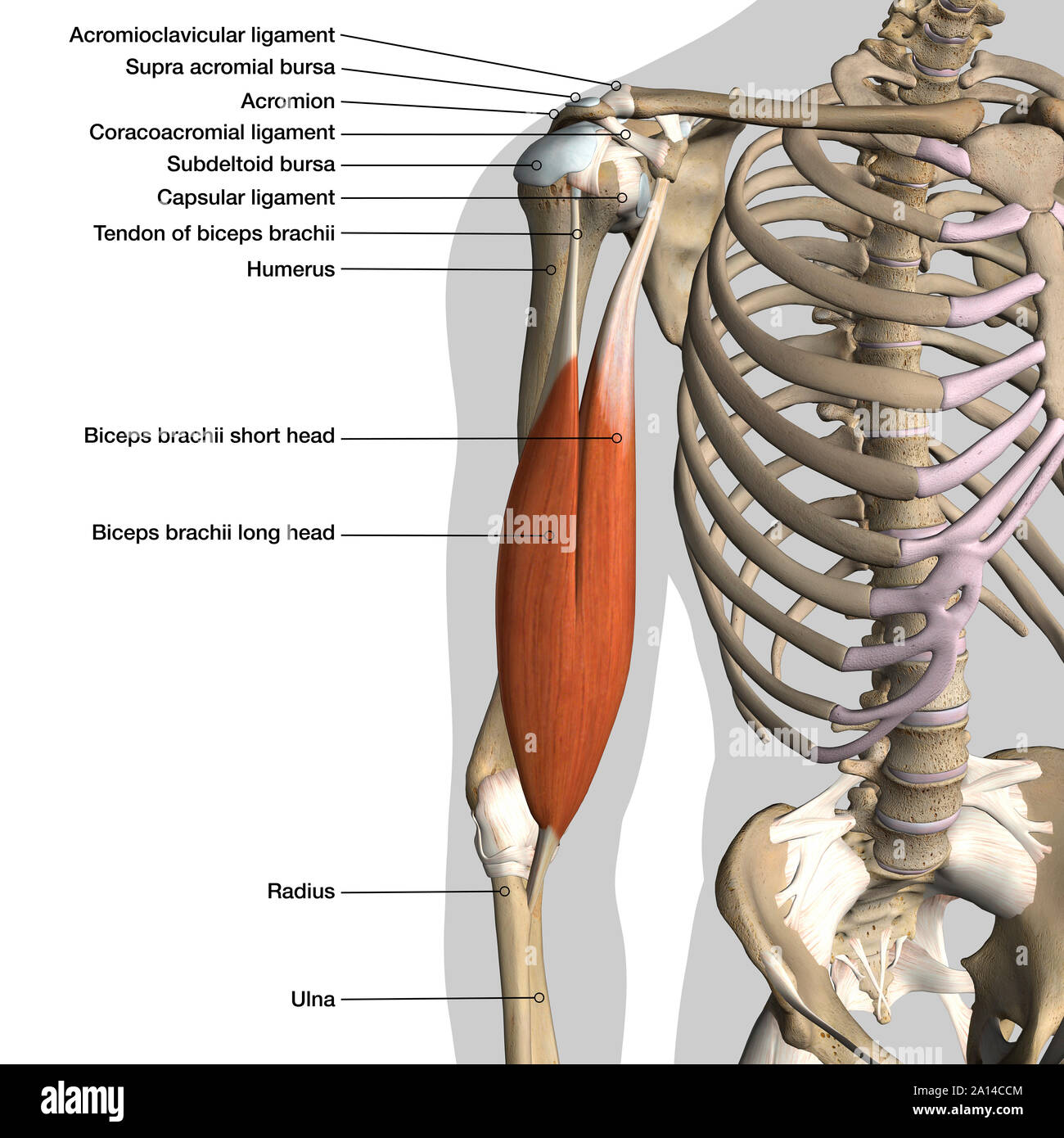
Labeled anatomy chart of male biceps muscle and shoulder ...

Anatomy Pregnant Vector & Photo (Free Trial) | Bigstock
Abdominal muscles diagram hi-res stock photography and images ...
Muscles of the Abdominal Wall - ppt video online download

Found on google search engine I typed anatomy and physiology ...

11.4 Identify the skeletal muscles and give their origins ...
:focal(749x0:751x2)/GettyImages-1192965863-2000-e1583791850721-ead103f609e84db993821e671abe1b1c.jpg)
The Complete Guide to Your Abs Muscles

Abdominal Muscles Photograph by Microscape/science Photo ...
Post a Comment for "45 abdominal muscles labeled"