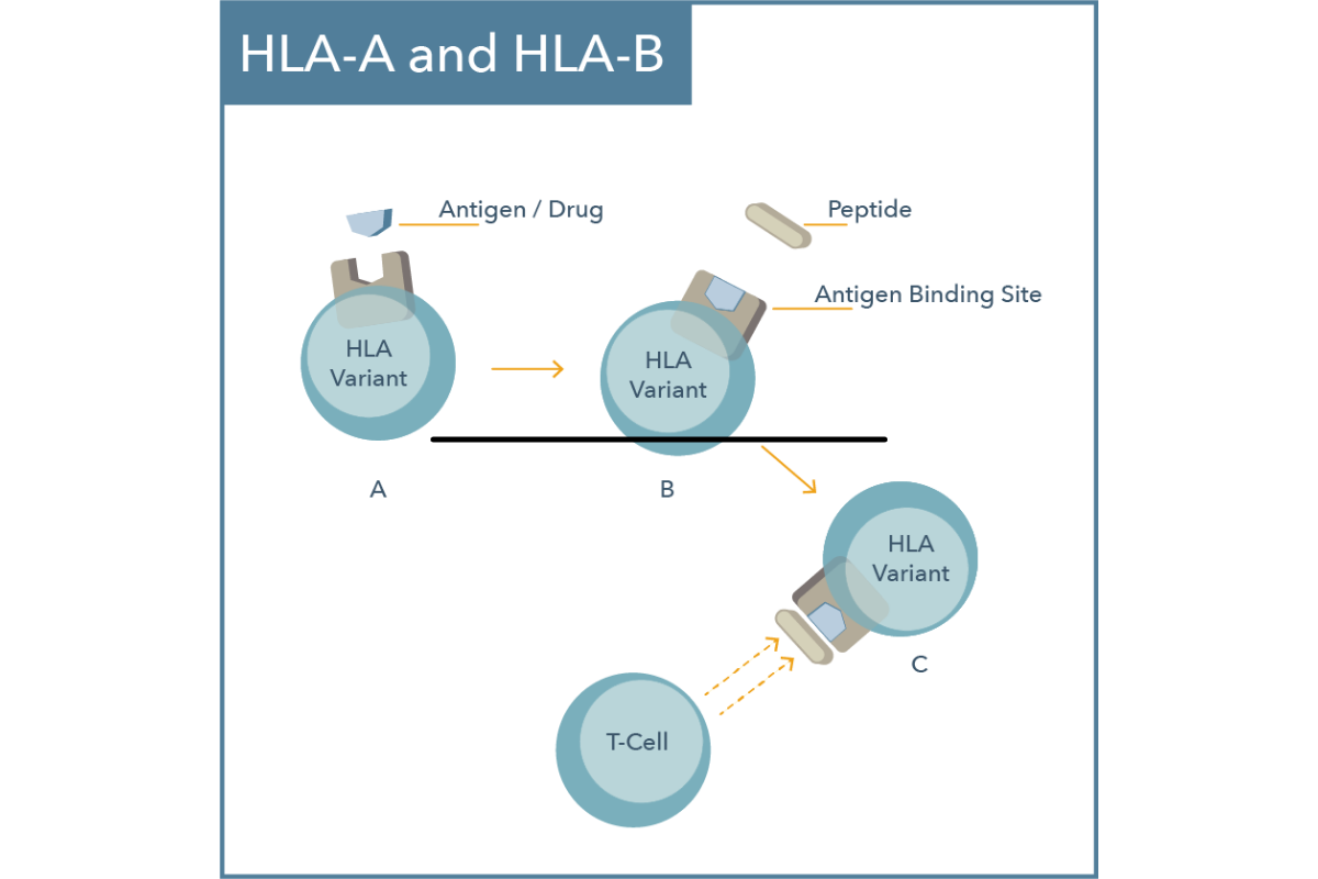
42 which label or labels indicate(s) the antigen binding site?
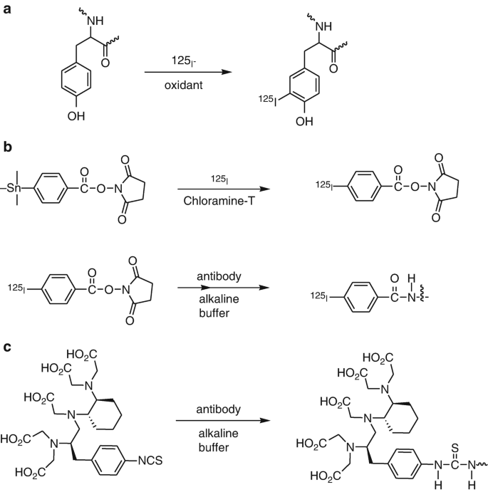
M A&P Introduction to Lymphatic System and Immunity Label the cause of infection and some structures involved in fighting the infection. After the proteins are separated by electrophoresis, the _____. ... Which label or labels indicate(s) the antigen binding site? the two sites labeled with the letter A. Steps in antigen presentation include which of these? Overview of Protein Labeling | Thermo Fisher Scientific - US Overview of Protein Labeling. Biological research often requires the use of molecular labels that are covalently attached to a protein of interest to facilitate detection or purification of the labeled protein and/or its binding partners. Labeling strategies result in the covalent attachment of different molecules, including biotin, reporter ...
Multiplex label-free biosensor for detection of ... - ScienceDirect The labels may destroy the binding sites that participate in antibody-antigen interactions, on the one hand, and non-specifically interact with proteins immobilized on a surface, on the other hand (Cooper, 2009; Fan et al., 2008; Orlov et al., 2018; Seydack, 2005). The label-free techniques are not prone to the label-associated issues.

Which label or labels indicate(s) the antigen binding site?
(PDF) Kuby immunology 7th Ed | Tabitha Royal - Academia.edu chapter 1 I Historical Perspective I Innate Immunity I Adaptive Immunity I Comparative Immunity I Immune Dysfunction and Its Consequences Numerous T Lymphocytes ... Antibody Types: IgM, IgA, IgD, IgG, IgE and Camelid Antibodies However, since pentameric IgM has 10 antigen binding sites, it has higher avidity (overall binding strength) for antigens than IgG and acts as an excellent activator of the complement system and ... Anatomy and Physiology 2: Chapter 22: Immune System Indicate whether the label identifies a specific or nonspecific form of defense. Specific: B-lymphocytes, antibodies, agglutinin, complement (antibody-dependent pathway), major histocompatability complexes, cytotoxic T-cells, memory T-cells, plasma cells, immunoglobulin, T-lymphocytes, antigen presenting cells, helper T-cells, CD4+ cells
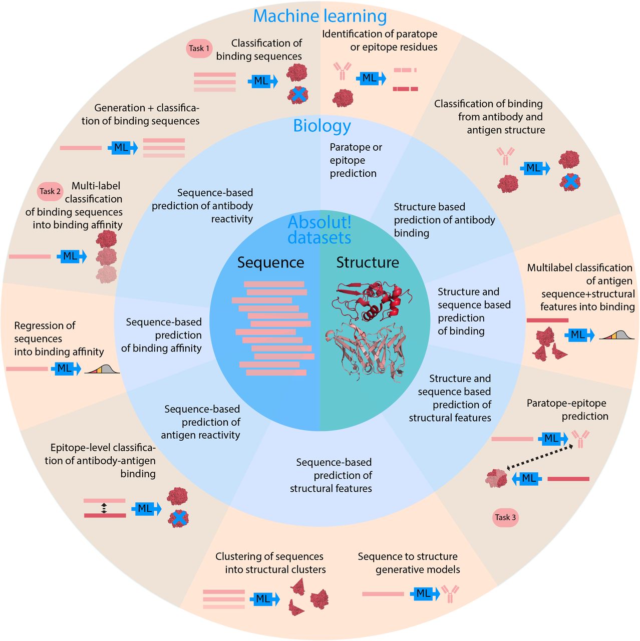
Which label or labels indicate(s) the antigen binding site?. Antibody Labeling - an overview | ScienceDirect Topics Double labeling of cells indicates the presence RALDH2 as a synthetic enzyme ... were labeled with monoclonal antibodies to pore-forming protein and bound ... Learning context-aware structural representations to predict antigen ... our framework comprises three components to leverage biological insights: (i) graph convolutions to capture the spatial relationships of the interfaces, (ii) an attention layer to enable each protein's interface predictions to account for the potential binding context provided by its partner and (iii) transfer learning to leverage the larger set … Immunoassay - Wikipedia In addition to the binding of an antibody to its antigen, the other key feature of all immunoassays is a means to produce a measurable signal in response to the binding. ... Most, though not all, immunoassays involve chemically linking antibodies or antigens with some kind of detectable label. A large number of labels exist in modern ... The structure of a typical antibody molecule - NCBI Bookshelf by CA Janeway Jr · 2001 · Cited by 103 — An antibody is identical to the B-cell receptor of the cell that secretes it ... giving an antibody molecule two identical antigen-binding sites (see Fig.
Exam 3: Mastering Lymphatic System (#6) Flashcards | Quizlet Which of these statements about lymphocytes is false? -They bind antigens. -They mostly occur in lymphoid tissues. -They are phagocytic. -They occur as B, T, and NK types. they are phagocytic Classes of lymphocytes T cells B cells NK cells T cells are approximately ___% of circulating lymphocytes. 80% B cells are ___% of circulating lymphocytes. Antibody Structure - University of Arizona An antigenic determinant, a site on the antigen that the immune system responds to by making antibody, can frequently be one unique structure on the antigen. In hen egg white lysozyme, a glutamine at position 121 (Gln 121) protrudes away from the antigen surface. In this view, Gln 121 is circled. The antibody is not shown. Fluorescent Labeling of Antibody Fragments Using Split GFP Antibody fragments are easily isolated from in vitro selection systems, such as phage and yeast display. Lacking the Fc portion of the antibody, they are usually labeled using small peptide tags recognized by antibodies. In this paper we present an efficient method to fluorescently label single chain Fvs (scFvs) using the split green fluorescent protein (GFP) system. A 13 amino acid tag ... A Developmentally Regulated Kinesin-related Motor Protein ... The same K7 fragment that was used for the generation of a K7/MBP antigen (residues 1–520) was cloned into the EcoRI site of the histidine tag expression vector pET23a (Novagen, Madison, WI). The coding sequence of the Aequorea victoria green fluorescent protein (S65T GFP mutant; Heim et al. , 1995 ) was cloned into the Hin cII and Xho I ...
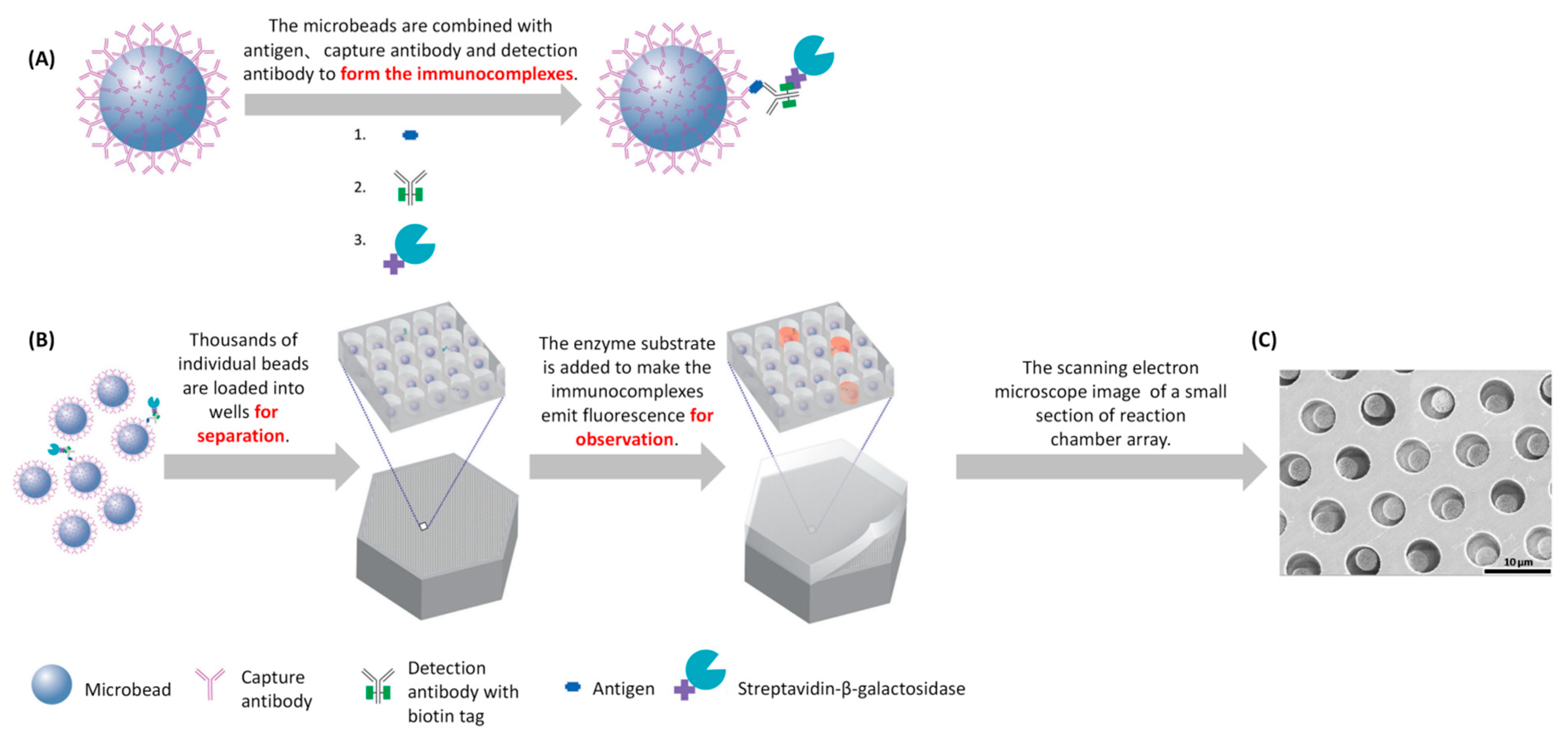
Novel non-nucleic acid targets detection strategies based on ... Notably, various non-nucleic acid targets are also biomarkers of environmental pollution, human disease and prognosis and food contamination. Traditional methods for detecting non-nucleic acid targets include high-performance liquid chromatography (HPLC), gas chromatography (GC), mass spectrometry (MS), magnetic resonance imaging (MRI), enzyme-linked immunosorbent assay (ELISA), etc. Anatomy Labs for Exam 2 Flashcards | Quizlet Which label or labels indicate(s) the antigen binding site? The two sites labeled with the letter A. What defense mechanism is shown in the images above? Complement (group of proteins that work together, binding to antibodies and forming large holes in target cells) What defense mechanism is shown in this image? Destruction by an NK cell. Concatenated Metallothionein as a Clonable Gold Label for Electron ... Whether indirect or direct, labelling of the protein may be sparse because of inaccessibility of the tag to its target or because of low affinity of the tag for its target. Moreover, other sites may be labeled because of adventitious binding of the tag to other proteins (Jahn 1999; Hainfeld and Powell 2000). Together, these difficulties point ... Immunoassay of total prostate-specific antigen using europium(III ... In two-site immunoassays, europium (III) chelate-labeled SA is typically utilized for the binding of biotinylated antibodies and signal generation. The indirect labeling of SA with europium (III) chelate-labeled BSA-polypeptide (Glu:Lys) conjugate has enabled further improvements in assay sensitivity.
Exam 2 Lab Flashcards | Chegg.com Which label or labels indicate (s) the antigen binding sight? The two sides labeled D The two sides labeled A The single location labeled B The two sites labeled C A What defense mechanism is shown in the images above? Complement Pyrogen Interferon Perforin Complement What defense mechanism is shown in this image?
Antibody Conjugation One method to reduce the number of undesirable conjugations is the replacement of cysteine with selenocysteine. This reduces the probability of conjugation occurring at inappropriate sites of the polypeptide chain(s) that disrupt the structure of the antibody (and therefore its affinity and/or specificity) [].In the case of antibody-drug conjugates, modification of glutamine residues using the ...
A&P2 Lab 10 HW Flashcards | Quizlet Study with Quizlet and memorize flashcards containing terms like Drag the labels onto the diagram to identify the lymphoid tissues and organs of the lymphatic system., Drag the labels onto the diagram to identify the structural features of the spleen., Which of the labels indicates a structure through which lymph flows? and more.
Dissecting the treatment-naive ecosystem of human melanoma ... Jul 07, 2022 · This suggests that antigen presentation alone is not sufficient to promote anti-tumor immunity, as recent work in patient models suggests (Ho et al., 2022), and may indicate areas prone to cancer immune evasion, possibly through expression of TIMP1 (Zurac et al., 2016).
PDF Secondary - Novus Bio Variable domain heavy chain Contains antigen binding site, confers antibody specificity (H+L) Heavy + light Whole immunoglobulin (heavy and light chain) α Alpha heavy chain IgA class δ Delta heavy chain IgD class ε Epsilon heavy chain IgE class γ Gamma heavy chain IgG class µ Mu heavy chain IgM class κ Kappa light chain λ Lambda light chain
Different Ways to Add Fluorescent Labels - Thermo Fisher Scientific Labeling various targets with separate fluorescent colors allows you to visualize different structures or proteins within a cell in the same experiment. Ways to fluorescently label your target include fluorescent dyes, immunolabeling, and fluorescent fusion proteins —all of which can provide a means to selectively mark structures and proteins ...
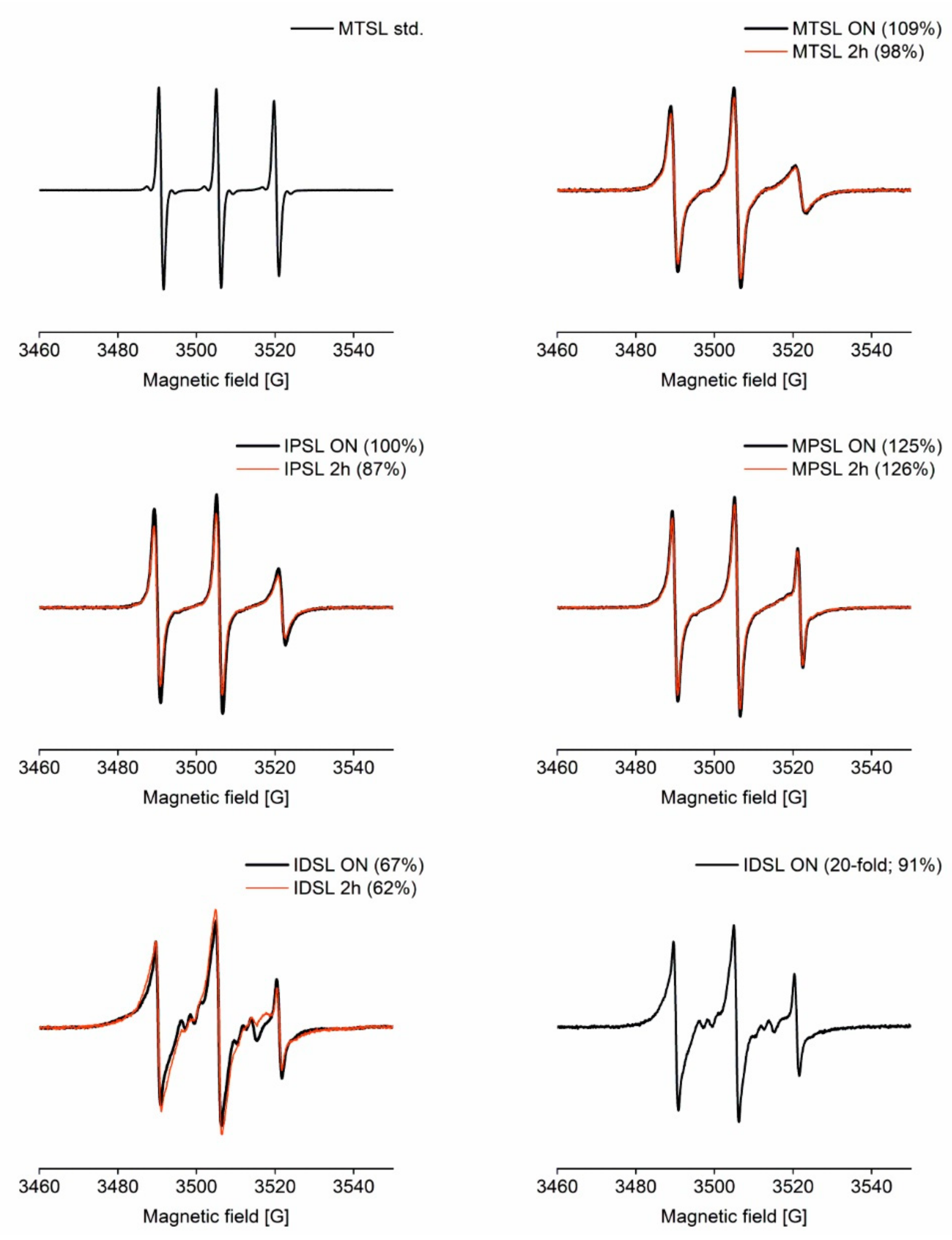
Conformation of peptides bound to the transporter associated with ... For resolving the structure of bound peptides and the mechanism of peptide binding, we used EPR spectroscopy in combination with site-directed spin labeling of antigenic peptides to ( i) probe the spatial arrangement of the peptide-binding pocket of TAP and ( ii) to elucidate the conformation of the bound peptide. Go to: Results
Why Site-Specific — AlphaThera Site-specific antibody labeling ensures that the label does not interfere with antigen binding. This becomes particularly important when attaching large biomolecules to the antibody (e.g. enzymes), which can sterically prevent antigen binding. Site-specific labeling also ensures the proper orientation of immobilized antibodies.
News - Default | Packaging Connections India’s largest multinational in flexible packaging materials & solutions and a global leader in polymer sciences has been riding the waves of innovation to build packaging products, applications & solutions that will further enhance the role of packag
Western Blot Technique: Principle, Steps, Uses | Microbe Online Sep 17, 2022 · This will prevent the non-specific binding of the antibody and reduce the overall background signal. Common blocking buffers include 5% non-fat dry milk or BSA in a TBS-Tween solution. However, do not use a milk solution when probing with phosphor-specific antibodies as it can cause high background from its endogenous phosphoprotein, casein.
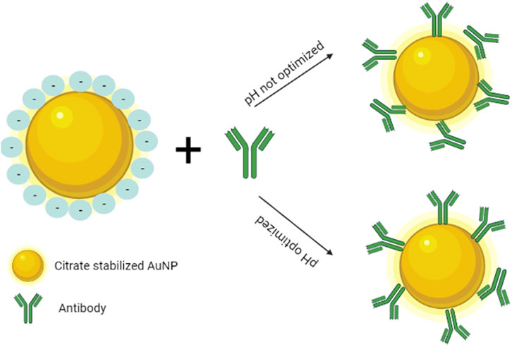
Lateral flow assays: Principles, designs and labels An alternative biorecognition molecule of antibody is immunogen, which is a cheaper biomolecule than an antibody, since the production cost of antibody is high. The binding of antibody to colloidal gold occurs by means of hydrophobic residues in the antigen-binding site of antibody. This way, the binding sites of an antibody are decreased.
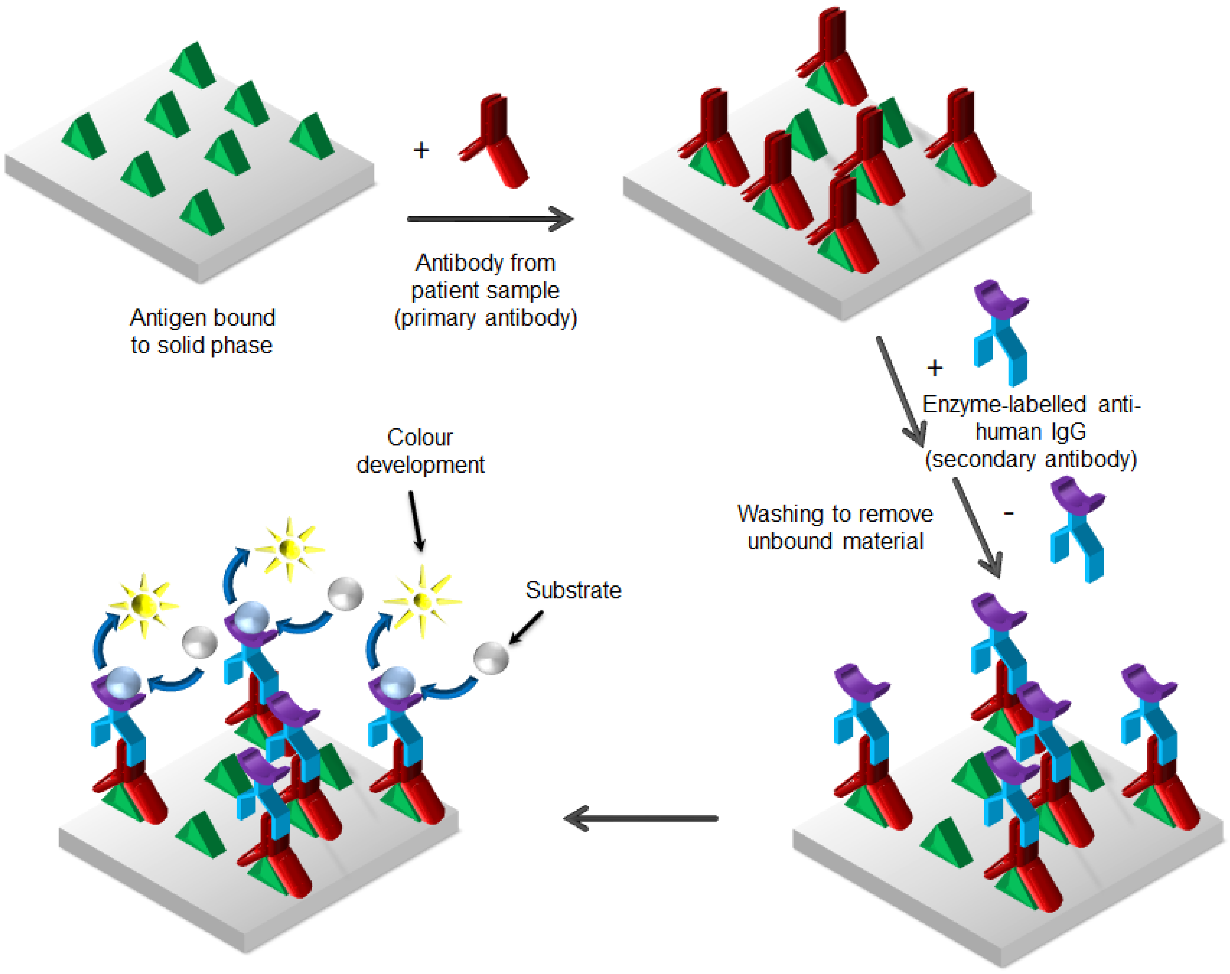
EIAs and ELISAs | Microbiology | | Course Hero A red color (from gold particles) or blue (from latex beads) developing at the test line indicates a positive test. If the color only develops at the control line, the test is negative. Like ELISA techniques, lateral flow tests take advantage of antibody sandwiches, providing sensitivity and specificity.
Fluorescence - Wikipedia Fluorescence is the emission of light by a substance that has absorbed light or other electromagnetic radiation.It is a form of luminescence.In most cases, the emitted light has a longer wavelength, and therefore a lower photon energy, than the absorbed radiation.
Structure and Function of Immunoglobulins - PMC - PubMed Central (PMC) The antigen binding site is the product of a nested gradient of diversity (A) H chain rearrangement can yield as many as 38,000 different VDJ combinations. The addition of nine N nucleotides on either side of the D gene segment yields can yield up to 64,000,000 different CDR-H3 junctional sequences.
Immunolabeling | Thermo Fisher Scientific - US For direct immunofluorescence, the antibody binding the epitope is labeled with fluorophores (green). For indirect or secondary detection, the primary antibody binds the epitope and a fluorophore-labeled secondary antibody (purple) that has specificity for the primary antibody binds to it. Direct immunofluorescence
Structure of Antibody (With Diagram) | Organisms - Biology This variable region, composed of 110-130 amino acids, gives the antibody its specificity for binding antigen. ADVERTISEMENTS: Antibodies are of five classes - IgG, IgA, IgM, IgD and IgE. Ig stands for immunoglobulins. IgG constitutes to about 75% of the total antibodies. IgE is involved in allergy and IgM is formed during the primary response.
Real-Time, label-free monitoring of tumor antigen and serum antibody ... The binding kinetics and affinity values of anti-NY-ESO-1 monoclonal antibody, ES121, to the cancer-testis antigen NY-ESO-1 were determined according to the surface heterogeneity model and resulted in K D values of 1.3×10 −9 and 2.1×10 −10 M. The reconfigured instrument was then used to measure the interaction between tumor antigens and ...
Abzymes: Catalytic Antibodies - UC Santa Barbara Rotate around active site complex. Label charged or partially charged centers. Labels off. For best results, push the buttons in sequence. 1. Zoom into the antigen-binding site with bound hapten. Initially, the ... The germline sequences of the heavy chain gene segments indicate that this particular asparagine residue arose as the result of ...
HW 29.docx - Best of Home Work (Exercise 29: Blood) Drag... Label the red blood cells with the correct antigen (s). Left to right… a antigen, b antigen, a & b, none Left to right … a antigen , b antigen , a & b , none Antibodies are proteins that have a lock-and-key recognition for their antigen established by the antigen-binding site on the antibody.
Anatomy and Physiology 2: Chapter 22: Immune System Indicate whether the label identifies a specific or nonspecific form of defense. Specific: B-lymphocytes, antibodies, agglutinin, complement (antibody-dependent pathway), major histocompatability complexes, cytotoxic T-cells, memory T-cells, plasma cells, immunoglobulin, T-lymphocytes, antigen presenting cells, helper T-cells, CD4+ cells
Antibody Types: IgM, IgA, IgD, IgG, IgE and Camelid Antibodies However, since pentameric IgM has 10 antigen binding sites, it has higher avidity (overall binding strength) for antigens than IgG and acts as an excellent activator of the complement system and ...
(PDF) Kuby immunology 7th Ed | Tabitha Royal - Academia.edu chapter 1 I Historical Perspective I Innate Immunity I Adaptive Immunity I Comparative Immunity I Immune Dysfunction and Its Consequences Numerous T Lymphocytes ...
















Post a Comment for "42 which label or labels indicate(s) the antigen binding site?"