44 neuron structure labeled
Neuron Anatomy Diagram | Quizlet Start studying Neuron Anatomy. Learn vocabulary, terms, and more with flashcards, games, and other study tools. Neuron under Microscope with Labeled Diagram - AnatomyLearner The neuron structure has two main components: the cell body and the neuron processes (axons and dendrite). Let's see the neuron histology slide labelled diagram and try to find out the below-mentioned characteristics - Presence of an identifiable cell body (soma) that locates in the brain's grey matter (according to the slide image).
Parts of a Neuron and How Signals are Transmitted Neurons are the basic building blocks of the nervous system. These specialized cells are the information-processing units of the brain responsible for receiving and transmitting information. Each part of the neuron plays a role in communicating information throughout the body.

Neuron structure labeled
A Labelled Diagram Of Neuron with Detailed Explanations They are found in the brain, spinal cord and the peripheral nerves. A neuron is also known as the nerve cell. The structure of a neuron varies with their shape and size and it mainly depends upon their functions and their location. Neurons are the structural and functional units of the nervous system. A group of neurons forms a nerve. Neuron Structure and Classification Structural classification of neurons. 1) Bipolar; 2) Multipolar and 3) Unipolar. Bipolar neurons have only two processes that extend in opposite directions from the cell body. One process is called a dendrite, and another process is called the axon. Although rare, these are found in the retina of the eye and the olfactory system. Neuron Diagram Unlabeled Tactile Neuron Diagram ( Unlabeled) by trynne is licensed under the Creative Commons.Neuron Anatomy Activity The parts of the neuron have been labeled. Your challenge is to write the correct name for each part and explain what it does. Between the axon ending and the dendrite of the next neuron is a very tiny gap called the synapse (or synaptic ...
Neuron structure labeled. Single-neuron projectome of mouse prefrontal cortex | Nature ... Mar 31, 2022 · Prefrontal cortex (PFC) is the cognitive center that integrates and regulates global brain activity. However, the whole-brain organization of PFC axon projections remains poorly understood. Using ... Types of Neurons: Parts, Structure, and Function - Verywell Health Interneurons are the most abundant neurons in the body. They act as the signal controllers within the body, relaying important information from one end of the nervous system to the other. The interneurons sit in the middle of other neurons, such as motor or sensory neurons. They are responsible for relaying electrical signals. Molecular dynamics simulation for all - PMC 19.09.2018 · Neuron 54, 511–533 [PMC free article] [Google Scholar] Mirjalili V, and Feig M (2013). Protein Structure Refinement through Structure Selection and Averaging from Molecular Dynamics Ensembles. Journal of Chemical Theory and Computation 9, 1294–1303. [PMC free article] [Google Scholar] Mobley DL, and Dill KA (2009). Pyramidal cell - Wikipedia Ramón y Cajal was also the first person to propose the physiological role of increasing the receptive surface area of the neuron. The greater the pyramidal cell's surface area, the greater the neuron's ability to process and integrate large amounts of information. Dendritic spines are absent on the soma, while the number increases away from it.
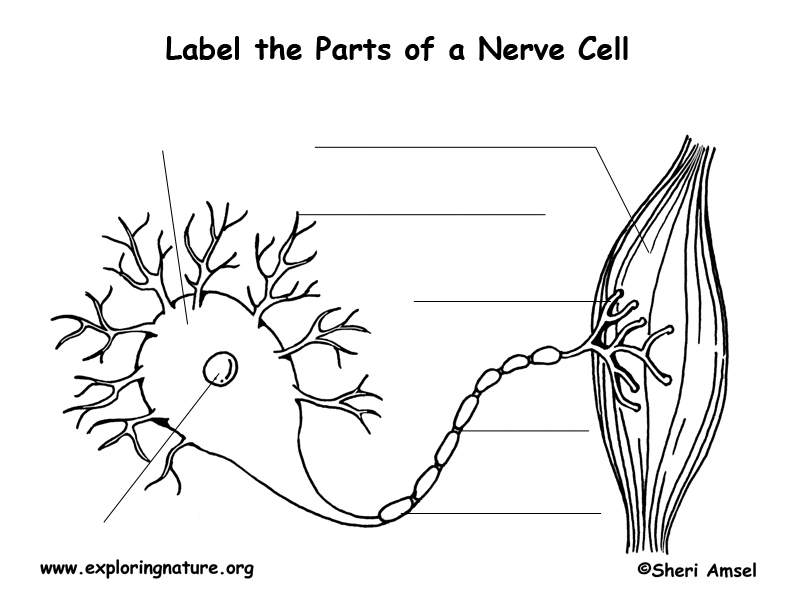
Label Neuron Anatomy Printout - EnchantedLearning.com Read the definitions, then label the neuron diagram below. axon - the long extension of a neuron that carries nerve impulses away from the body of the cell. cell body - the cell body of the neuron; it contains the nucleus (also called the soma) dendrites - the branching structure of a neuron that receives messages (attached to the cell body) Labelled Diagram Of Motor Neuron - schematron.org on Labelled Diagram Of Motor Neuron. These receive incoming impulses from other neurones. Diagram of a nerve cell A motor neurone. Where two neurones meet there is a small gap called a. Find Motor neuron, detailed and accurate, labeled Stock Vectors and millions of other royalty-free stock photos, illustrations, and vectors in the Shutterstock. Motor Units in Skeletal Muscle - GetBodySmart 11.12.2017 · Here’s how to learn all muscles with quizzes and labeled diagrams. 1. 2. When a motor neuron fires, all the muscle fibers in the motor unit contract at once. The size of a motor unit varies from just a few fibers in the eye muscles (precise movements) to over a thousand fibers in the large leg muscles (powerful movements). Micrograph demonstrating a … A Labelled Diagram of Neuron with Detailed decription A neuron is a type of cell that is largely responsible for conveying information via electrical and chemical impulses. The brain, spinal cord, and peripheral nerves all contain them. The nerve cell is another name for a neuron. The structure of a neuron changes depending on its form and size, as well as its function and location.
Location, Structure, and Functions of Motor Neurons - Bodytomy Being the most basic units of the human nervous system, neurons play a vital role in sensing and responding to different external as well as internal stimuli. A motor neuron is one of the three types of neurons involved in this process. Read about the structure and function of a motor neuron with reference to a neatly labeled diagram, in this Bodytomy post. What Is a Neuron? Diagrams, Types, Function, and More Neurons usually have one main axon. Dendrites Dendrites are fibrous roots that branch out from the cell body. Like antennae, dendrites receive and process signals from the axons of other neurons.... Overview of neuron structure and function - Khan Academy The peripheral nervous system ( PNS ), which consists of the neurons and parts of neurons found outside of the CNS, includes sensory neurons and motor neurons. Sensory neurons bring signals into the CNS, and motor neurons carry signals out of the CNS. _Image modified from " Nervous system diagram ," by Medium69 ( CC BY-SA 4.0 )._ Medium spiny neuron - Wikipedia Medium spiny neurons (MSNs), also known as spiny projection neurons (SPNs), are a special type of GABAergic inhibitory cell representing 95% of neurons within the human striatum, a basal ganglia structure. Medium spiny neurons have two primary phenotypes (characteristic types): D1-type MSNs of the direct pathway and D2-type MSNs of the indirect pathway.
Exploration of the Human Spinal Cord
What Is a Neuron? - Definition, Structure, Parts and Function Axon is a tube-like structure that carries electrical impulse from the cell body to the axon terminals that passes the impulse to another neuron. Synapse It is the chemical junction between the terminal of one neuron and dendrites of another neuron. Also Read: Difference between neurons and neuroglia Neuron Types

Zaf Naqui | Anatomy
Neuroanatomy, Motor Neuron - StatPearls - NCBI Bookshelf Jul 31, 2021 · While the term “motor neuron” evokes the idea that there is only one type of neuron that conducts movement, this is far from the truth. In fact, within the classification of a “motor neuron,” there lies both upper and lower motor neurons, which are entirely different in terms of their origins, synapse points, pathways, neurotransmitters, and lesion characteristics. Overall, motor ...
Nerve Cell (Neuron) Labeling Page
Nervous System - Label the Neuron Name: Choose the correct names for the parts of the neuron. (1) (2) (3) (4) (5) (6) This neuron part receives messages from other neurons. (7) This neuron part sends on messages to other neurons. (8) This neuron part gives messages to muscle tissue. (9) This neuron part processes incoming messages. (10)

Nervous tissue
Single-neuron projectome of mouse prefrontal cortex - Nature 31.03.2022 · To relate the branching structure of neural trees with its developmental history, we implemented a scheme to order segments for each neuron. First, we assigned the axon path from the soma to a ...
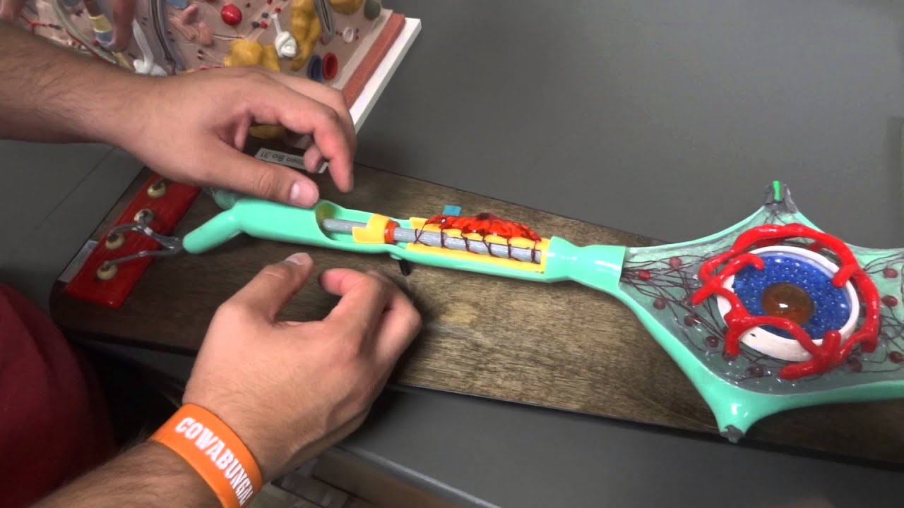
Axon/Neuron Model Anatomy - YouTube
Muscular System Labeled Diagram Stock Photos, Pictures ... Contraction synapse vector illustration. Labeled medical structure scheme. Contraction synapse vector illustration. Labeled closeup medical structure scheme. Diagram with full cycle of ACH release, action potential, troponin bonding and filament muscular system labeled diagram stock illustrations

Light-Up Neuron
Diagram Quiz on Neuron Structure and Function (Labeling Quiz) 4. Name the cell type, labeled '3' that produce the myelin sheath around neuronal axon Schwann cell glial cell Axonites neurons 5. The organelles are embedded in the viscous fluid region of the neuron labeled '4' is Soma nucleus cytoplasm axoplasm 6. The region labeled '5 ', is also called as soma, that contains the nucleus and cytoplasm Nucleus
Structure of the Spinal Cord, Reflexes, and Nerves Week #12 Flashcards ...
Neuron Structure - 3D Models, Video Tutorials & Notes - AnatomyZone Neurons are the basic structural and functional unit of the nervous system, whereas glial cells play a supportive role. Cell Body Just like any other cell in the body, the cell body of the neuron contains the nucleus, and other major organelles. So within the nucleus, you've got chromosomal DNA, and you also might have a small nucleolus.

Axon/Neuron Anatomy Model - YouTube
Histology of neurons: Morphology and types of neurons | Kenhub Cerebellum - molecular, Purkinje, granular layers. Peripheral nerves - epineurium, perineurium, endoneurium. This article will explain the histology of neurons, providing you with information about their structure, types, and clinical relevance. It will also cover briefly the histological layers of the central and peripheral nervous systems.
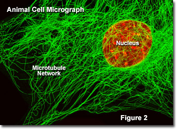
Molecular Expressions Cell Biology: Microtubules
Location, Structure, and Functions of Motor Neurons - Bodytomy A motor neuron is one of the three types of neurons involved in this process. Read about the structure and function of a motor neuron with reference to a neatly labeled diagram, in this Bodytomy post. Motor Neuron Disease (MND) A motor neuron disease affects the normal functioning of motor neurons, resulting in their degeneration and death.
Post a Comment for "44 neuron structure labeled"